The College of Sciences boasts a wide variety of facilities on and off campus that provide students with resources and environments necessary for academic success. The information below allows you to get better acquainted with the facilities and laboratories available to science students.


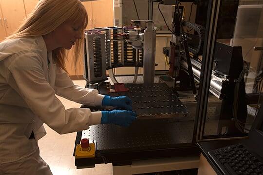
Electron Microanalysis & Imaging Laboratory (EMiL)
The Electron Microanalysis & Imaging Laboratory is an analytical laboratory dedicated to the solution of problems using electron micro-beam techniques for clients in industry, government agencies, and academia. EMiL is capable of providing microanalysis and imaging services.
Genomics Core Facility
The Genomics Core Facility aids in the development and support of genomics-based research. It provides equipment and training for DNA microarray handling, Western blot/gel/microarray scanning, PCR amplification, and analysis of DNA, RNA, and protein samples. The facility is located in the Science and Engineering Building, Rooms 3122, 3123, and 3124.
Las Vegas Isotope Science Laboratory (LVIS)
The Las Vegas Isotope Science Laboratory is equipped for analyses of water and geologic and biologic samples for oxygen, hydrogen, carbon, and nitrogen stable isotopes. These stable isotopes are powerful tracers of natural processes, ranging widely from tracking of bird migrations to studies of trophic levels and paleoclimate.
Nevada Extreme Conditions Lab (NEXCL)
The Nevada Extreme Conditions Lab (NEXCL) is a multidisciplinary group that explores fundamental experimental, computational, and engineering problems of materials under high pressure.
Nevada Isotope Geochronology Laboratory (NIGL)
The Nevada Isotope Geochronology Laboratory offers facilities to conduct 40Ar/39Ar dating. The geoscience department operates mineral separation facilities. The department also has a hydrology research group that focuses on surface and groundwater modeling projects.
Science and Engineering Building (SEB)
The Science and Engineering Building, a state-of-the-art building, is a unique environment for interdisciplinary research and education on the UNLV campus. The building’s innovative design supports collaboration among faculty in the sciences, engineering, health sciences, and other university research units.
Confocal and Biological Imaging Core
The UNLV Confocal and Biological Imaging Core is equipped with three advanced microscopy platforms that, together, support deep‑tissue multiphoton imaging, high‑sensitivity and enhanced‑resolution confocal imaging, and lifetime‑based multicolor experiments for fixed and live samples.
-
Nikon A1R MP multiphoton/confocal: Multiphoton ("Two-Photon") and confocal laser‑scanning system with a tunable femtosecond pulsed dual‑input infrared laser (including a Coherent Discovery Dual Beam 1300nm IR laser for multiphoton imaging) and high‑speed resonant scanner, enabling rapid, low‑photodamage time‑lapse imaging deep within thick or living specimens such as brain slices, tissues, and intravital preparations.
-
Zeiss LSM 800 with Airyscan: Point‑scanning confocal microscope equipped with four solid‑state laser lines and GaAsP detectors plus an Airyscan super‑resolution module that delivers approximately 1.7‑ to 1.8‑fold higher resolution and improved signal‑to‑noise for dim samples, ideal for quantitative 3D imaging of fixed cells, tissue sections, and gentle live‑cell experiments.
-
Leica Stellaris with WLL and TauSense: Confocal platform with a tunable pulsed white‑light laser, 405 nm diode, and five HyD detectors providing prism‑based spectral detection up to approximately 850 nm, combined with TauSense lifetime tools and LIGHTNING deconvolution for enhanced‑resolution multicolor imaging and functional FLIM‑based contrast; an Oko UNO stage-top CO₂ mixer and incubation system for extended live mammalian cell imaging, and fast z‑correction support long‑term live‑cell, tissue, and organoid imaging as well as advanced analyses such as spectral unmixing, FRET, FRAP, and FLIP.
Wesley E. Niles Herbarium
Wesley E. Niles Herbarium is a collection of dried, pressed plant specimens mounted on archival-quality sheets of paper that are placed in folders and stored in cabinets. These specimens provide tangible documentation for the occurrence, distribution, and diversity of flowering plants, conifers, and mosses in the Southwestern desert regions of the United States, especially the Mojave Desert.